Venous Interventions
Catheter-directed DVT thrombolysis
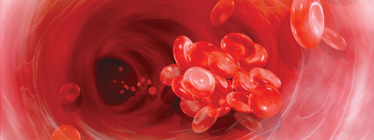
Deep vein thrombosis (DVT) is a blood clot in a vein, most often in the leg. Left untreated, it can lead to leg pain, swelling, leg ulcers and even the loss of mobility. Over time, the clot can break up and travel to the lungs, where it can lead to a serious condition called pulmonary embolism (PE).
Catheter-directed DVT thrombolysis is a minimally invasive procedure to treat DVT. An interventional radiologists guides a small catheter to the site of the DVT, where a special drug is administered directly into the clot, effectively dissolving it to remove the danger to the patient.
DVT thrombolysis has been shown to completely break down DVT blood clots more effectively than anticoagulation medication, as well as maintain better blood flow within the veins. Risks are minimal, and it can help to minimize future scarring of the veins from DVT. It is an outpatient procedure, and may require a 1-2 night hospital stay depending on the age of the clot.
Our interventional radiologists also specialize in chronic DVT (clots that are more than 2 months old). We also treat May-Thurner disease, including stenting of the iliac vein.
Inferior vena cava filter placement/removal (including complex extra caval filters)
An IVC filter is a small filter that is placed within the inferior vena cava, the large vein that transports blood back to the heart from the legs. It effectively “traps” clots within it, causing them to break up into smaller and less threatening pieces while also allowing blood to flow around it. IVC filters may be placed on a permanent or temporary basis to decrease the risk of pulmonary embolism.
During the procedure, an interventional radiologist uses imaging to guide a catheter into the inferior vena cava that contains the filter. A contrast material is injected into the vein to help assess the ideal location. The IVC filter is then placed in the correct position, where it expands to affix itself to the walls of the blood vessel.
If the placement of the IVC filter is not permanent, a future procedure to retrieve the filter will be necessary. This will happen when the threat of a pulmonary embolism has passed.
As a minimally invasive procedure, no surgical incisions are necessary. IVC filters have a high success rate when it comes to protecting patients from a pulmonary embolism. It is also an effective treatment for patients who are not able to control clots from forming with medication. Risks are rare, and include the potential for infection and an allergic reaction to the contrast material that is used to help position the filter.
In some cases, if an IVC filter remains in the body too long, it can move or fracture within the vein. AMIC interventional radiologists have specialized experience with the complex retrieval of fractured, embedded and penetrating IVC filters.
Varicose Veins and Spider Veins
Varicose veins are very common; about half of all adults aged 40-69 have them. That equates to about 20 million people in the United States who are suffering from varicose veins or spider veins, which appear on the surface and give the skin a discolored appearance. Varicose veins are the result of a venous insufficiency (also known as venous reflux), a condition resulting from decreased blood flow from the leg veins to the heart, and which causes blood to “pool” within the veins, causing them to bulge.

